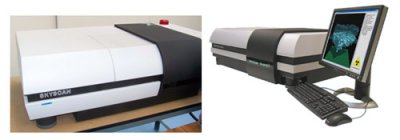
A private laboratory specialized in the analysis, testing and failure analysis of materials since 1993
A private laboratory specialized in the analysis, testing and failure analysis of materials since 1993

Principle
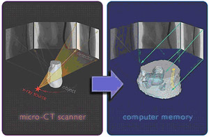
The X-beam passes through the sample, and is then collected by a detector. The X-ray intensity loss between the source and the detector is linked to the atomic structure of the material, that permits to draw the X-Rays radiography (like a medical X-Rays radiography).
The 2D sample rotation gives different X-Rays pictures (different angles). All those pictures are then collected and re-built to form a 3D reconstruction. Thus the inner structure of the sample can be 3D-observed and inspected.
Characteristics :
![]() X-ray source ................ 20-100kV,10W,<5µm spot size
X-ray source ................ 20-100kV,10W,<5µm spot size
![]() X-ray detector ..............11Mp, 12-bit cooled CCD fiber-optically coupled to scintillator
X-ray detector ..............11Mp, 12-bit cooled CCD fiber-optically coupled to scintillator
![]() Maximum object size... 27mm in diameter(single scan) or 50mm in diameter (offset scan)
Maximum object size... 27mm in diameter(single scan) or 50mm in diameter (offset scan)
![]() Detail detectability ...... <0.8µm at highest resolution
Detail detectability ...... <0.8µm at highest resolution
![]() Radiation safety ......... <1µSv / h at any point on the instrument surface
Radiation safety ......... <1µSv / h at any point on the instrument surface

Featured accessories :
Cryogenic freezing system : Ice block observation (sample holder on the left, and 3D-reconstructed sample on the right)
Micro-positioning sample holder : to improve the sample centering